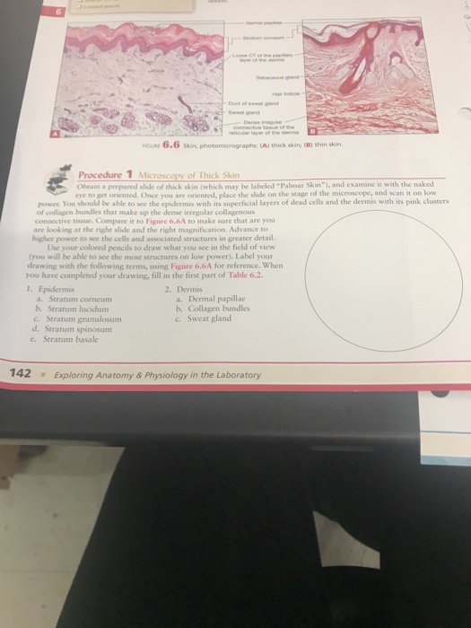
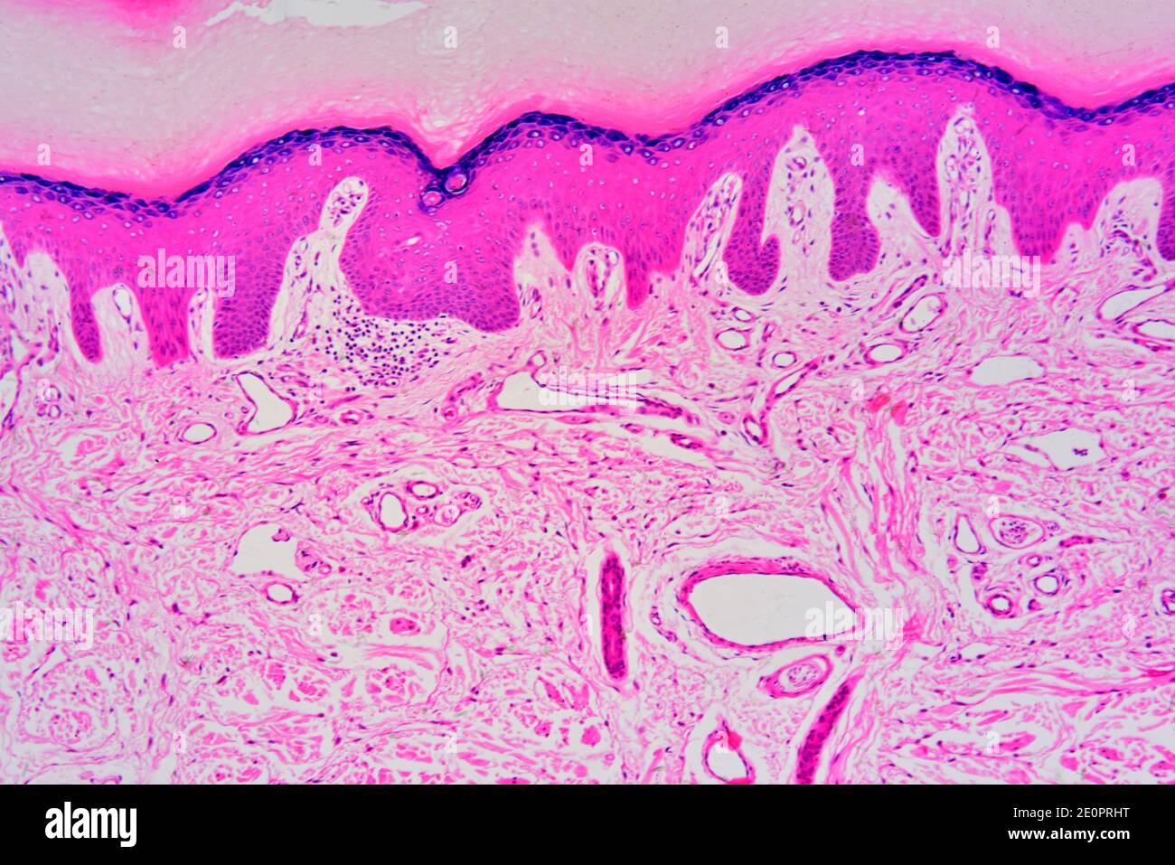
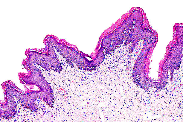
39 label the photomicrograph of thin skin.
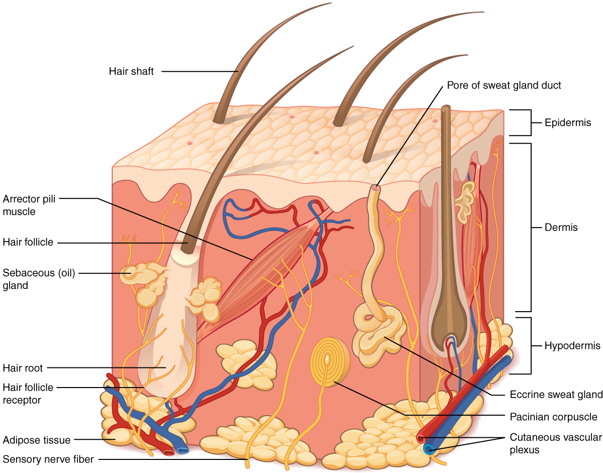
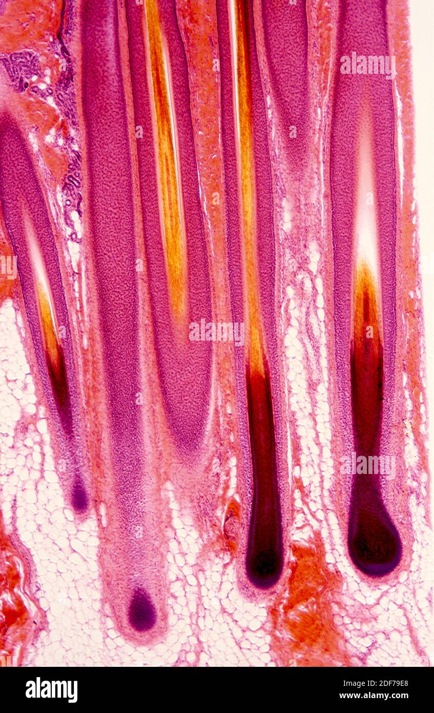
Final Exam A&P 1 Flashcards | Quizlet Label the photomicrograph of thin skin Hair shaft, epidermis, dermal root sheath, sebaceous gland, dermis, hair matrix label the structures of the hair follicle Identify the layers of the epidermis with relation to their location and role in keratinization ... the receptors responsible for olfaction are located in the olfactory epithelium Figure 7.4 Photomicrograph of the skin and accessory ... Papilla of hair a projection of connective tissue into the hair follicle and contains blood vessels that provide nutrients to the dividing cells of the matrix. Sebaceous Gland Oil glands that surround hair follicles; secrete oils that lubricates skin, hair, and into the neck of the hair follicle. Hair Follicle
photomicrographs of thin skin Flashcards | Quizlet photomicrographs of thin skin. Term. 1 / 4. stratum corneum. Click the card to flip 👆.

Label the photomicrograph of thin skin.
Layers of the Skin | Anatomy and Physiology I - Lumen Learning Skin that has four layers of cells is referred to as “thin skin.”. From deep to superficial, these layers are the stratum basale, stratum spinosum, stratum granulosum, and stratum corneum. Most of the skin can be classified as thin skin. “Thick skin” is found only on the palms of the hands and the soles of the feet. (Solved) - Label the photomicrograph of thin skin. Label the ... Aug 5, 2021 · Label the photomicrograph of thin skin 1 answer below » Label the photomicrograph of thin skin 1 Approved Answer Mohinee k answered on August 05, 2021 4 Ratings ( 9 Votes) Label the photomicrograph of the skin A photograph taken with the help of microscope . Skin is the largest sensory organ in body.its protect the body sense pain... solution .pdf Figure 7.1: Photomicrograph of Skin Diagram | Quizlet Start studying Figure 7.1: Photomicrograph of Skin. Learn vocabulary, terms, and more with flashcards, games, and other study tools.
Label the photomicrograph of thin skin.. Solved Label the photomicrograph of thin skin. Dermis Duct ... Question: Label the photomicrograph of thin skin. Dermis Duct of sebaceous gland Hair Follicle Sebaceous gland Hair Epidermis This problem has been solved! See the answer Show transcribed image text Expert Answer 100% (37 ratings) A … View the full answer Transcribed image text: Label the photomicrograph of thin skin. Figure 7.1: Photomicrograph of Skin Diagram | Quizlet Start studying Figure 7.1: Photomicrograph of Skin. Learn vocabulary, terms, and more with flashcards, games, and other study tools. (Solved) - Label the photomicrograph of thin skin. Label the ... Aug 5, 2021 · Label the photomicrograph of thin skin 1 answer below » Label the photomicrograph of thin skin 1 Approved Answer Mohinee k answered on August 05, 2021 4 Ratings ( 9 Votes) Label the photomicrograph of the skin A photograph taken with the help of microscope . Skin is the largest sensory organ in body.its protect the body sense pain... solution .pdf Layers of the Skin | Anatomy and Physiology I - Lumen Learning Skin that has four layers of cells is referred to as “thin skin.”. From deep to superficial, these layers are the stratum basale, stratum spinosum, stratum granulosum, and stratum corneum. Most of the skin can be classified as thin skin. “Thick skin” is found only on the palms of the hands and the soles of the feet.











Post a Comment for "39 label the photomicrograph of thin skin."