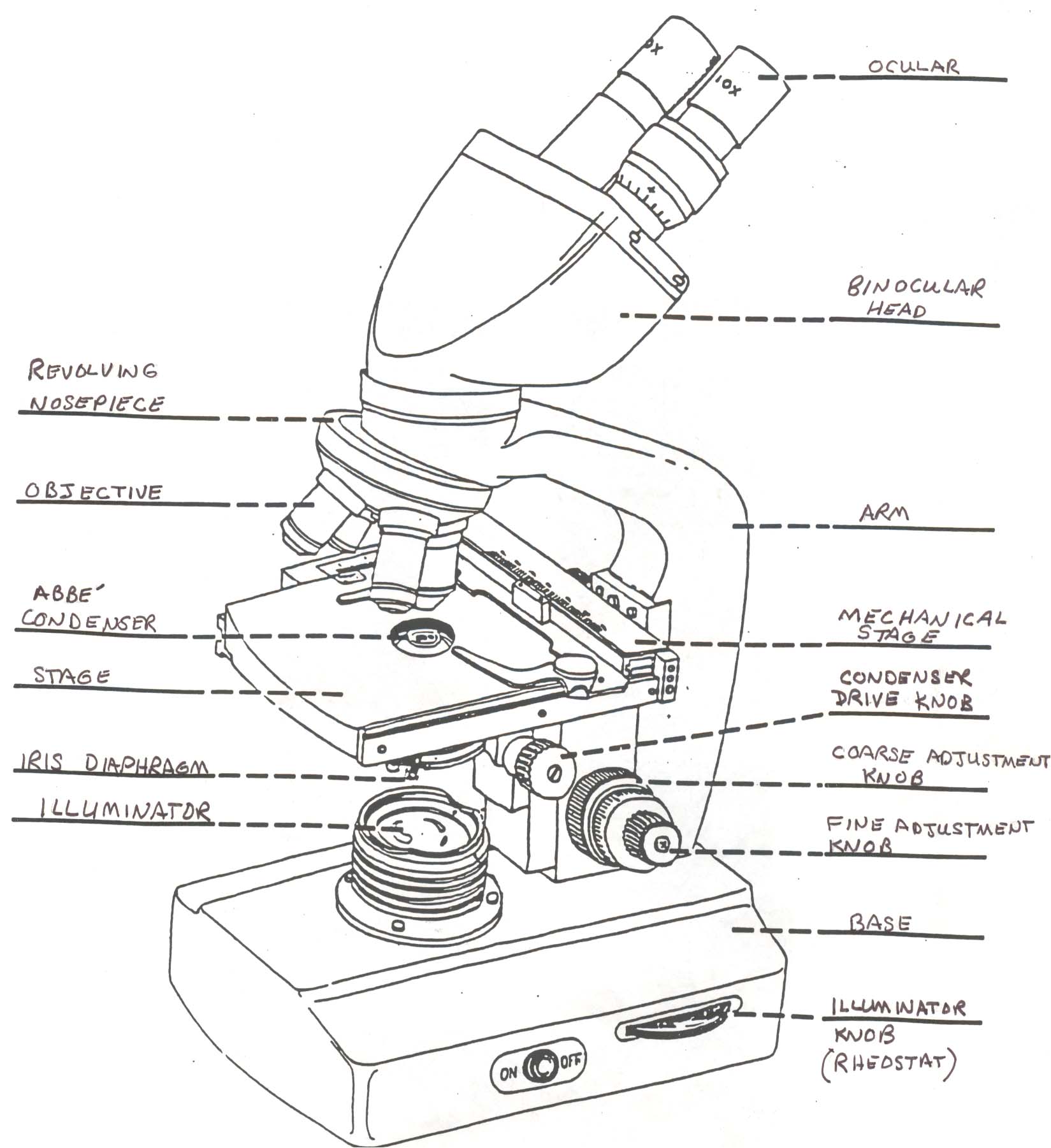
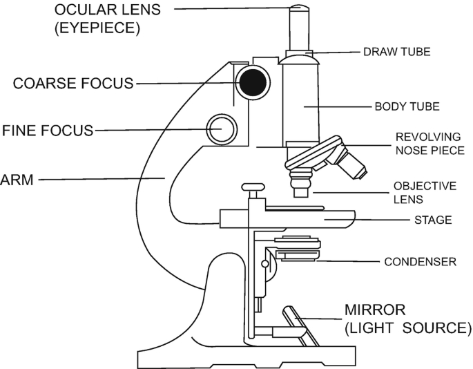
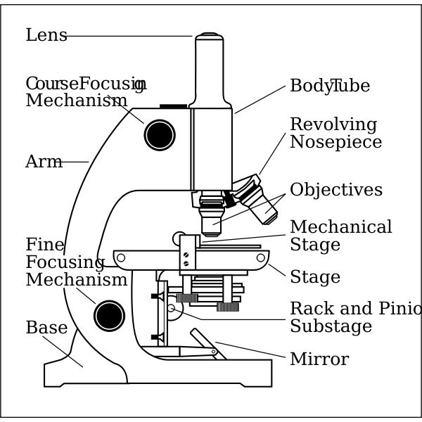
39 microscope drawing with label
How To Draw A Microscope - YouTube How To Draw A Microscope 135,831 views Aug 31, 2020 1.1K Dislike Share Art for Kids Hub 6.16M subscribers Today, we're learning how to draw a cool microscope! 👩🎨 JOIN OUR ART HUB MEMBERSHIP! Microscopy - Wikipedia Optical or light microscopy involves passing visible light transmitted through or reflected from the sample through a single lens or multiple lenses to allow a magnified view of the sample. The resulting image can be detected directly by the eye, imaged on a photographic plate, or captured digitally.The single lens with its attachments, or the system of lenses and imaging equipment, …
Microscope Drawing: How to Sketch Microscope Slides How to Draw Microscope Slides Organize and orient your field of view: To begin, draw a circle as large as possible with a pencil. An 8.5 x 11-inch piece of paper is good size for beginners. The circle represents what you see through the eyepiece of the microscope. Using thin lines, divide the circle into quarters in order to organize the picture.
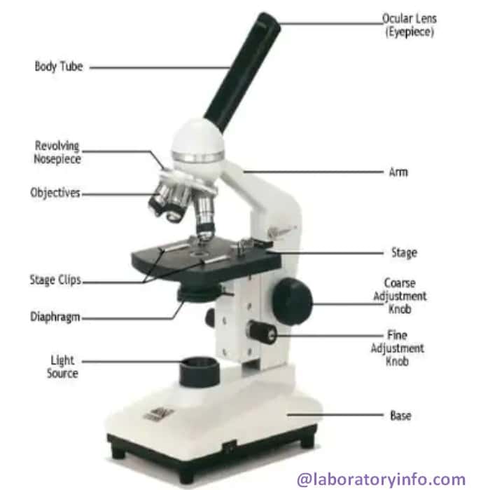
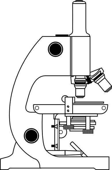
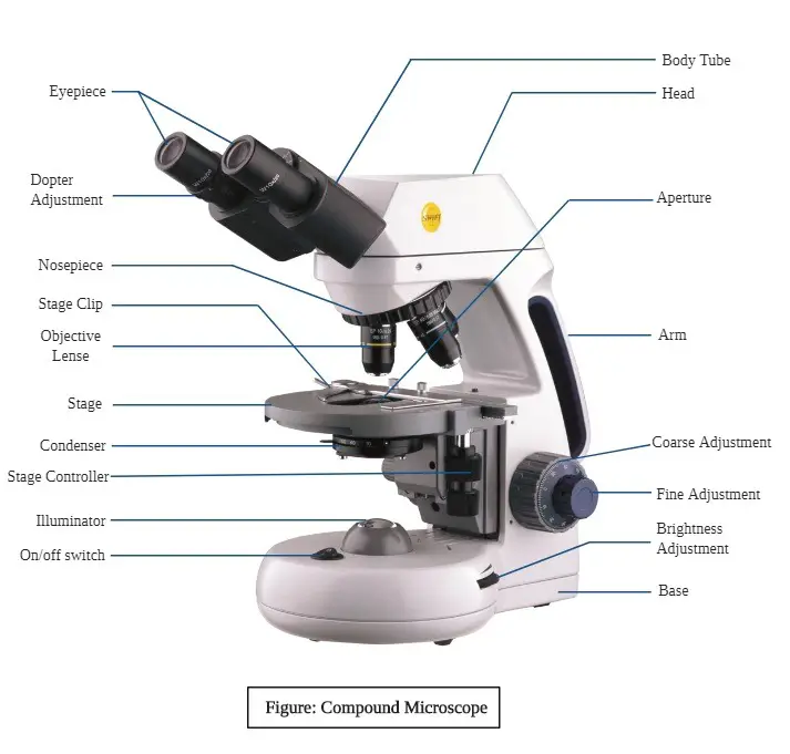
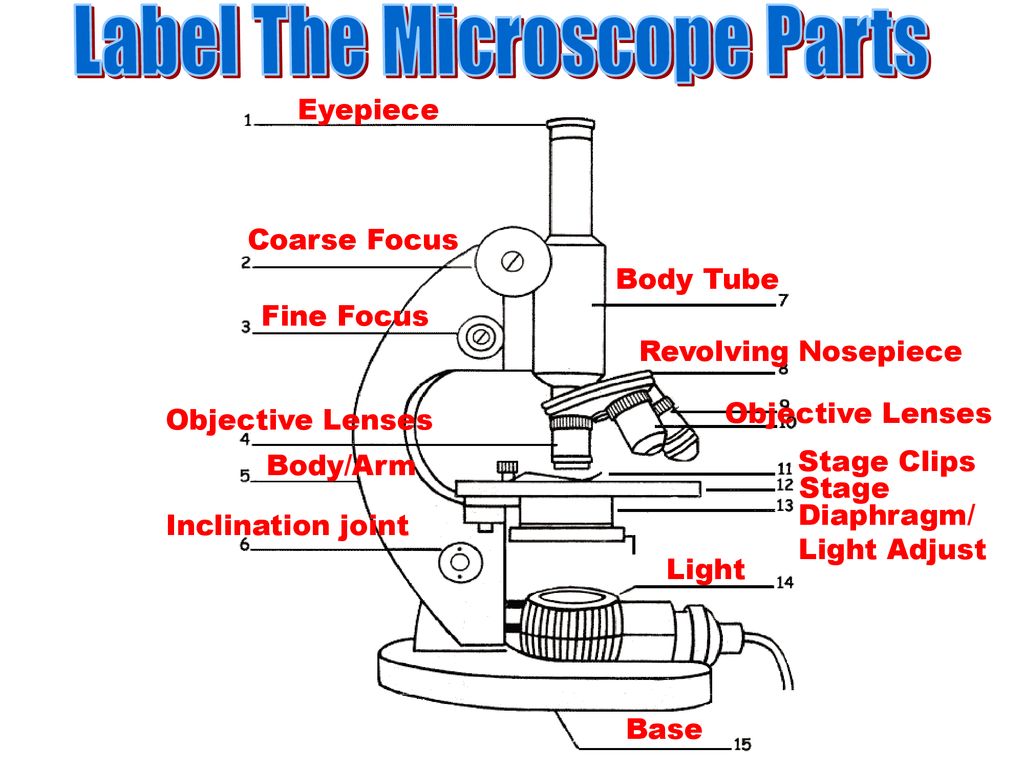
Microscope drawing with label
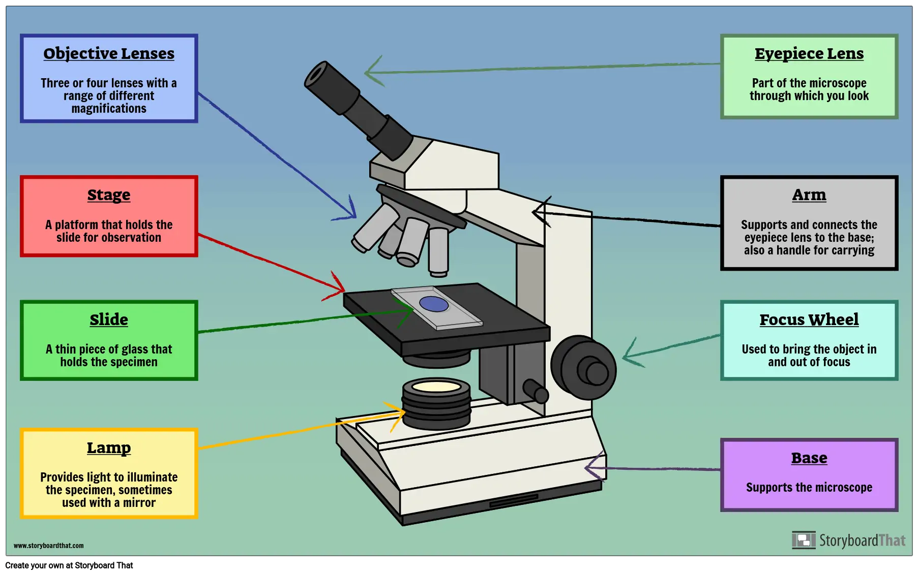
Microscope Labeling Diagram | Quizlet Focus and magnify light in differing amounts to view the specimen. Stage Clips. Hold the slide in place on the stage. Nosepiece. Holds the objective lenses and allows the lenses to rotate for viewing. Stage. Supports the slide where the specimen is being viewed. Lamp. Projects or reflects light upward through the diaphragm. Microscopes | Biomedx Take your health practice endeavors in biological microscopy to the moon (and the bank) with a Biomedx configured Olympus microscope system. With over two decades of implementing the best live blood / live cell qualitative auditing systems providing big screen, HD, and superior imaging capabilities, we know exactly what works and works best for practitioners; from the health coach through to ... How to Sketch a Microscope Slide - Identifying and Sketching Cell ... First, to represent the microscope field of view, draw a circle on the page - this can be freehand or, if you want to be precise, use a compass. If you are using a graticule slide (a microscope slide with millimeter grid lines), lightly sketch a grid over your circle.
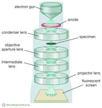
Microscope drawing with label. Microscope Imaging Software | Products | Leica Microsystems Aug 20, 2021 · Microscope Imaging Software. Microscope imaging software from Leica Microsystems combines microscope, digital camera and accessories into one fully integrated solution. With an intuitive user interface and straightforward navigation, it guides the user through any workflow, whether fast image acquisition or sophisticated expert analysis. Parts of the Microscope with Labeling (also Free Printouts) Parts of the Microscope with Labeling (also Free Printouts) By Editorial Team March 7, 2022 A microscope is one of the invaluable tools in the laboratory setting. It is used to observe things that cannot be seen by the naked eye. Table of Contents 1. Eyepiece 2. Body tube/Head 3. Turret/Nose piece 4. Objective lenses 5. Knobs (fine and coarse) 6. A Study of the Microscope and its Functions With a Labeled Diagram ... These labeled microscope diagrams and the functions of its various parts, attempt to simplify the microscope for you. However, as the saying goes, 'practice makes perfect', here is a blank compound microscope diagram and blank electron microscope diagram to label. Parts of a microscope with functions and labeled diagram - Microbe Notes Parts of a microscope with functions and labeled diagram April 19, 2022 by Faith Mokobi Having been constructed in the 16th Century, Microscopes have revolutionalized science with their ability to magnify small objects such as microbial cells, producing images with definitive structures that are identifiable and characterizable.
Online Auctions - Park Village contents of office 4: multi tier industrial shelving, datamax label printers x 4, 4 drawer steel cabinet x 2, steel racking x 3, assorted gears, 2 door small steel cabinet x 1, platform scale x 1, assorted plastic rolls/wrappers/bags, electric fans x 2 (subject to confirmation) estimate closing time : 2022-08-03 11:54:14 Compound Microscope Parts - Labeled Diagram and their Functions Labeled diagram of a compound microscope Major structural parts of a compound microscope There are three major structural parts of a compound microscope. The head includes the upper part of the microscope, which houses the most critical optical components, and the eyepiece tube of the microscope. UD Virtual Compound Microscope - University of Delaware ©University of Delaware. This work is licensed under a Creative Commons Attribution-NonCommercial-NoDerivs 2.5 License.Creative Commons Attribution-NonCommercial-NoDerivs 2.5 … Compound Microscope - Diagram (Parts labelled), Principle and Uses See: Labeled Diagram showing differences between compound and simple microscope parts Structural Components. The three structural components include. 1. Head. This is the upper part of the microscope that houses the optical parts. 2. Arm . This part connects the head with the base and provides stability to the microscope.
Gram Stain Technique - Amrita Vishwa Vidyapeetham 28.08.2022 · Drawing a circle on the underside of the slide using a glassware-marking pen may be helpful to clearly designate the area in which you will prepare the smear. You may also label the slide with the initials of the name of the organism on the edge of the slide. Care should be taken that the label should not be in contact with the staining reagents. Part 3: Preparation of the … Microscope Drawing Easy with Label - YouTube Microscope Drawing Easy with Label 886 views Apr 13, 2020 In this video I go over a microscope drawing that is easy with label. There is a blank copy at the end of the video to review on your own.... Label the microscope — Science Learning Hub In this interactive, you can label the different parts of a microscope. Use this with the ... 18,701 Microscope drawing Images, Stock Photos & Vectors - Shutterstock 18,701 microscope drawing stock photos, vectors, and illustrations are available royalty-free. See microscope drawing stock video clips Image type Orientation Color People Artists Sort by Popular Science Abstract Designs and Shapes College and University Art Styles Printing, Typography, and Calligraphy microscope line art chemistry laboratory
Microscope Parts and Functions Microscope Parts and Functions With Labeled Diagram and Functions How does a Compound Microscope Work? Before exploring microscope parts and functions, you should probably understand that the compound light microscope is more complicated than just a microscope with more than one lens.
Labelled Diagram of Compound Microscope The below mentioned article provides a labelled diagram of compound microscope. Part # 1. The Stand: The stand is made up of a heavy foot which carries a curved inclinable limb or arm bearing the body tube. The foot is generally horse shoe-shaped structure (Fig. 2) which rests on table top or any other surface on which the microscope in kept.
Drawing Of A Microscope And Label - Warehouse of Ideas Here presented 54+ microscope drawing and label images for free to download, print or share. Title Is Informative, Centered, And Larger Than Other Text. How to draw a microscope and label. Compound microscopes have furthered medical research, helped to solve crimes, and they have repeatedly proven invaluable in unlocking the secrets of the.
How to draw compound of Microscope easily - step by step I will show you " How to draw compound of microscope easily - step by step "Please watch carefully and try this okay.Thanks for watching.....#microscopedrawi...
Animal Cell Anatomy & Diagram - Enchanted Learning The cell is the basic unit of life. All organisms are made up of cells (or in some cases, a single cell). Most cells are very small; in fact, most are invisible without using a microscope. Cells are covered by a cell membrane and come in many different shapes. The contents of a cell are called the protoplasm. Glossary of Animal Cell Terms: Cell ...
Blueprint Flat File Cabinets, Map Cabinet, Flat Files | Drafting ... Customize and organize your file drawers. Drawer dividers for 5-Drawer Flat Files provide easy separation of materials. Dividers are self-sticking black 11 plastic sections that can easily be cut to accommodate your needs.
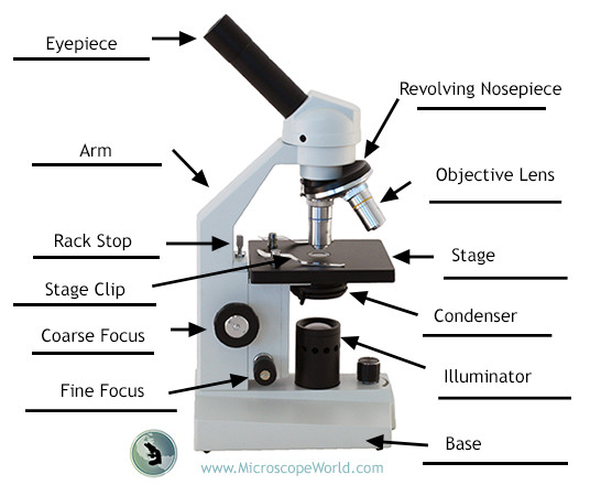
Compound Microscope Parts, Functions, and Labeled Diagram The individual parts of a compound microscope can vary heavily depending on the configuration & applications that the scope is being used for. Common compound microscope parts include: Compound Microscope Definitions for Labels Eyepiece (ocular lens) with or without Pointer: The part that is looked through at the top of the compound microscope. Eyepieces typically have a magnification between 5x & 30x.
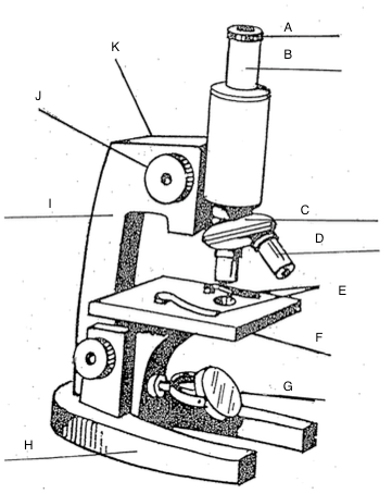
Label Microscope Diagram - EnchantedLearning.com Using the terms listed below, label the microscope diagram. arm - this attaches the eyepiece and body tube to the base. base - this supports the microscope. body tube - the tube that supports the eyepiece. coarse focus adjustment - a knob that makes large adjustments to the focus.
Cell Size and Scale - University of Utah Smaller cells are easily visible under a light microscope. It's even possible to make out structures within the cell, such as the nucleus, mitochondria and chloroplasts. Light microscopes use a system of lenses to magnify an image. The power of a light microscope is limited by the wavelength of visible light, which is about 500 nm. The most powerful light microscopes can …
Microscope Parts, Function, & Labeled Diagram - slidingmotion Microscope Parts Labeled Diagram The principle of the Microscope gives you an exact reason to use it. It works on the 3 principles. Magnification Resolving Power Numerical Aperture. Parts of Microscope Head Base Arm Eyepiece Lens Eyepiece Tube Objective Lenses Nose Piece Adjustment Knobs Stage Aperture Microscopic Illuminator Condenser Lens
COLLECTIBLES & DECORATIVE ARTS - Gardner Galleries Antique British brass microscope in mahogany case, unsigned but with R & J Beck objective, eyepiece, slides, fishplate and bullseye - the microscope measures 31.8cm (12 1/2in) high. Current Bid: $150. VIEW. 4836. ROLLUP DRAWING SET. Antique set in leather rollup case including rulers, dividers, pens, etc. Current Bid: $20. VIEW. 4837. TIFFANY STYLE LAMP. …
Microscope, Microscope Parts, Labeled Diagram, and Functions Microscope, Microscope Parts, Labeled Diagram, and Functions What is Microscope? A microscope is a laboratory instrument used to examine objects that are too small to be seen by the naked eye. It is derived from Ancient Greek words and composed of mikrós, "small" and skopeîn,"to look" or "see".
How to Sketch a Microscope Slide - Identifying and Sketching Cell ... First, to represent the microscope field of view, draw a circle on the page - this can be freehand or, if you want to be precise, use a compass. If you are using a graticule slide (a microscope slide with millimeter grid lines), lightly sketch a grid over your circle.
Microscopes | Biomedx Take your health practice endeavors in biological microscopy to the moon (and the bank) with a Biomedx configured Olympus microscope system. With over two decades of implementing the best live blood / live cell qualitative auditing systems providing big screen, HD, and superior imaging capabilities, we know exactly what works and works best for practitioners; from the health coach through to ...
Microscope Labeling Diagram | Quizlet Focus and magnify light in differing amounts to view the specimen. Stage Clips. Hold the slide in place on the stage. Nosepiece. Holds the objective lenses and allows the lenses to rotate for viewing. Stage. Supports the slide where the specimen is being viewed. Lamp. Projects or reflects light upward through the diaphragm.








![How To Draw A Microscope Step by Step - [12 Easy Phase]](https://easydrawings.net/wp-content/uploads/2021/01/Overview-for-Microscope-drawing.jpg)






Post a Comment for "39 microscope drawing with label"