43 correctly label the following anatomical features of the neuroglia
Cells of the Nervous System - Neurons - TeachMePhysiology Cell body or Soma - this contains the nucleus and the neurone's intracellular organelles (such as the mitochondria and Golgi apparatus). It is the center of neuronal metabolism. It also contains the Nissl Substance. These are granules containing rough endoplasmic reticulum and free ribosomes, making it the site of protein synthesis. Anatomy, Central Nervous System - StatPearls - NCBI Bookshelf The brain is an organ of nervous tissue that is responsible for responses, sensation, movement, emotions, communication, thought processing, and memory. Protection for the human brain comes from the skull, meninges, and cerebrospinal fluids. The nervous tissue is extremely delicate and can suffer damage by the smallest amount of force.
What Are Glial Cells and What Do They Do? - Verywell Health Your central nervous system (CNS) is made up of your brain and the nerves of your spinal column. Five types that are present in your CNS are: 1 Astrocytes Oligodendrocytes Microglia Ependymal cells Radial glia You also have glial cells in your peripheral nervous system (PNS), which comprises the nerves in your extremities, away from the spine.

Correctly label the following anatomical features of the neuroglia
Anatomy And Physiology Archive | July 04, 2021 | Chegg.com Question 1 (5 points) Match each of the following neuroglial cells to the best description of its function. myelination of axons in the CNS 1. astrocytes produce and circulate cerebrospinal fluid 2. e 1 answer a) Explain 'motion segment for human spine with the suitable diagram. 1 answer Diagnostics | Free Full-Text | Novel User-Friendly Application ... - MDPI such a user-based initialization was thus performed by a human user who was instructed to manually place the initial seed for each patient based on the following instructions: (i) the initial seed must be in the center of the cavity rather than on the border; (ii) the initial seed must be one of the darkest voxels in the cavity (i.e., have low … BIOL& 241 Human A & P 1 (6 credits) - Acalog ACMS™ 5. Draw and label the characteristic features of cardiac, skeletal, and smooth muscle tissues including the fascia associated with muscle tissue organization 6. Draw and label the characteristic features of a giant multipolar neuron and associated neuroglia 7. Identify designated bones and landmarks in the human skeleton 8.
Correctly label the following anatomical features of the neuroglia. Applied Sciences | Free Full-Text | A Novel Immersive Anatomy Education ... The Anat_Hub application offers a wide range of useful features for promoting active and self-regulated learning, such as 3D and AR modes, glossary, and quiz features. The application was tested with potential users, and on a variety of mobile device specifications. Very few volunteers have used AR prior to this study (13.2%). (Get Answer) - 6 Correctly label the following anatomical features of a ... 1. Sketch and label the following structures on a motor neuron smear: motor neuron (cell body, nucleus. nucleolus, dendrites, axon) and nuclei of neuroglial cells 2. Identify and label a sensory neuron cell body and a nerve fiber in a posterior... BIO 240 - Human Anatomy - Acalog ACMS™ Describe the tissue and cellular structures of the following: the respiratory bronchioles, the alveolar ducts, alveolar sacs, and the alveoli. Describe the structure of the respiratory membrane. Become familiar with the gross anatomy of the urinary system. Identify and locate the organs of the urinary system. Peripheral nervous system: Anatomy, divisions, functions | Kenhub Cranial nerves are peripheral nerves that mainly innervate anatomical structures of the head and neck. The exception to this is the vagus nerve, which also innervates various thoracic and abdominal organs. Cranial nerves originate from specific nuclei located in the brain.
Fundamentals Of Anatomy And Physiology Martini Pdf Free Download UPDATED The Eleventh Edition of Fundamentals of Anatomy & Physiology is a comprehensive textbook that fulfills the needs of today's students while addressing the concerns of their teachers. ... nosotros have made the following key changes: NEW SmartArt Videos help students better navigate primal, complex pieces of fine art. ... Hallmark Features of ... Nervous Tissue Structure And Function Worksheet Answers These new nerve cells or neurons and glial cells or glia Neurons are the basic functional units of the nervous system ring they generate electrical signals. What is a secondary organ is referenced... BIO 240 - Human Anatomy - Acalog ACMS™ A. Identify and locate the organs of the urinary system. B. Describe the tissues and regions of the organs of the urinary system. C. Relate the function of each of the organs of the urinary system with the gross structure. 6. Become familiar with the microscopic features of the kidney, the ureter, and the bladder. What Is a Cell Body? - Definition, Function & Types - Study.com The cell body, also called the soma, is the spherical part of the neuron that contains the nucleus. The cell body connects to the dendrites, which bring information to the neuron, and the axon ...
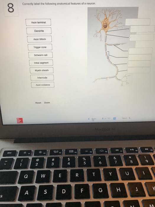
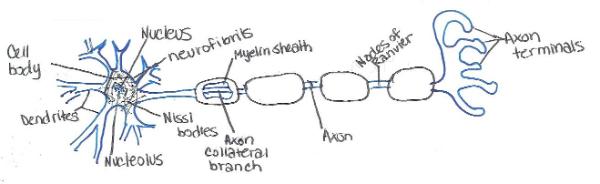
Correctly Label The Following Anatomical Features Of A Neuron First, the axon is a long, thin tube. Some of these branches are connected by a synapses called dendrites. In humans, the axon is over a meter long. Another important feature of a neuron is its synapse, a region of the plasma membrane that contains a single, continuous sarcomere. The nucleus of a neuron is large and round. archive.org › stream › NEW_1Full text of "NEW" - Internet Archive An icon used to represent a menu that can be toggled by interacting with this icon. PDF Neuron Worksheet Answers Just click on a worksheet, print it out and get to work. If the pages do not print out correctly from your computer setup, let me know: chudler@u.washington.edu Neuroscience for Kids - Worksheets Science worksheets: Label parts of a neuronStudents have to identify and label parts of a neuron (Axon, Nervous system: Structure, function and diagram | Kenhub Synonyms: Neuroglia Glial cells, also called neuroglia or simply glia, are smaller non-excitatory cells that act to support neurons. They do not propagate action potentials. Instead, they myelinate neurons, maintain homeostatic balance, provide structural support, protection and nutrition for neurons throughout the nervous system.
Complete Soccer Training: Overview of the nervous system
Human brain - Wikipedia The human brain is the central organ of the human nervous system, and with the spinal cord makes up the central nervous system.The brain consists of the cerebrum, the brainstem and the cerebellum.It controls most of the activities of the body, processing, integrating, and coordinating the information it receives from the sense organs, and making decisions as to the instructions sent to the ...

34 Correctly Label The Following Anatomical Features Of The Neuroglia ...
Neuromuscular Junction Structure and Functions - New Health Advisor The junction synapse has 3 characteristic features: There are two membranes called the pre and post synaptic membranes. There exists a distinct space between these membranes and is known as Synaptic Cleft. High density of small spherical vesicles are present, which contain neurotransmitter substances.
32 Correctly Label The Following Anatomical Features Of A Neuron ...
› 41956428 › COGNITIVE_NEUROSCIENCECOGNITIVE NEUROSCIENCE THE BIOLOGY OF THE MIND Fourth Edition Enter the email address you signed up with and we'll email you a reset link.

Neuronal Cell Anatomy in detail
RDP_ Tissues 2021 - slideshare.net Sketch and label the various types of major tissues from slides and identify their specific location and a specific function. "The body contains at least 200 distinct cell types. These cells contain essentially the same internal structures, yet they vary enormously in shape and function.

Schwann Cell Anatomy - Human Anatomy - GUWS Medical
catalog.jccc.edu › coursedescriptions › biolBiology (BIOL) < Johnson County Community College - JCCC a. Identify the elements of the skull and the major surface features of each. b. Classify the vertebrae by their regional characteristics. c. Identify the elements of the vertebral column and the major features of each. d. Identify the elements of the rib cage and their major surface features. e. Classify the articulated ribs as true, false or ...
Exercise 17: Histology of Nervous Tissue Flashcards | Easy Notecards
Cerebrum: Anatomy, Function, and Treatment - Verywell Health It is divided into two halves, or hemispheres, and its outer layer has large folds and creases of tissue that give the brain its characteristic wrinkly appearance. The cerebrum is responsible for processing sensory functions like vision, hearing, and touch; and it is involved in movement of your body.
Solved: Correctly Label The Following Anatomical Features ... | Chegg.com
quizlet.com › 445722440 › anatomy-midterm-lectureAnatomy Midterm Lecture Flashcards - Quizlet Correctly label the following anatomical features of the neuroglia. Correctly label the structures associated with unmyelinated nerve fibers in the PNS. Correctly label the following parts of a chemical synapse.

Solved: Correctly Label The Following Anatomical Features ... | Chegg.com
Neuromuscular Junction | Structure, Function, Summary & Clinical Neuromuscular junction is a microstructure present at the junction of motor neurons and the skeletal muscle fibers. It acts as a bridge connecting the skeletal system and the nervous system. The neuromuscular junction is a chemical synapse. The presynaptic terminal is the axonal terminal of. motor neuron containing synaptic vesicles.
Post a Comment for "43 correctly label the following anatomical features of the neuroglia"