42 nucleus unlabeled
Labeled Neuron Diagram | Science Trends The nucleus contains the neuron's DNA and is the site of RNA transcription and translation which produce proteins necessary for the neuron to function properly. The neuron transports proteins from the soma to the axon and dendrites via the activity of microtubule-associated motor proteins. Types of Neurons Live-dead assay on unlabeled cells using phase imaging with ... Feb 07, 2022 · The evolution of the cell dry mass and nucleus area for the labeled and unlabeled populations reveal that the chemical reagents decrease viability. The nondestructive approach presented here may...
Multiscale Assay of Unlabeled Neurite Dynamics Using Phase Imaging with ... In this work, we show that PICS can be used to measure the arborization process in unlabeled neural cultures, over multiple days, nondestructively (Figure 1). Our method consists of highly sensitive QPI as well as end-to-end image analysis to infer the fluorescence intensity for Tau and MAP2,41commonly used to identify axons and dendrites.
Nucleus unlabeled
Live-dead assay on unlabeled cells using phase imaging with ... Feb 07, 2022 · The evolution of the cell dry mass and nucleus area for the labeled and unlabeled populations reveal that the chemical reagents decrease viability. The nondestructive approach presented here may find a broad range of applications, from monitoring the production of biopharmaceuticals to assessing the effectiveness of cancer treatments. Neuron Diagram Unlabeled neuron, (1). axon, cell body, dendrites, nucleus, terminal. Unlabeled diagram of a motor neuron (try labeling: axon, dendrite, cell body, myelin, nodes of Ranvier, motor end plate).Read the definitions, then label the neuron diagram below. axon - the long extension of a neuron that carries nerve impulses away from the body of the cell. (PDF) Evidence of GABA-immunopositive neurons in the dorsal part of the ... Evidence of GABA-immunopositive neurons in the dorsal part of the lateral geniculate nucleus of reptiles: Morphological correlates with interneurons. Neuroscience, 1992. Jacques Repérant.
Nucleus unlabeled. Edinger-Westphal nucleus - Wikipedia From Wikipedia, the free encyclopedia The Edinger–Westphal nucleus (accessory oculomotor nucleus) is the parasympathetic pre-ganglionic nucleus that innervates the iris sphincter muscle and the ciliary muscle . Plant Cell Diagram Labels We Have got 13 images about Unlabeled Plant Cell Diagram images photos pictures backgrounds and more. ... visual instructional tool assists in grasping and retaining the names of the cell parts like mitochondrion vacuole nucleus and more with ease. It is a rigid covering made up of cellulose which a complex substance is providing structural ... Unlabeled Neuron Diagram - schematron.org Oct 20, 2018 · axon, cell body, dendrites, nucleus, terminal.The spinal cord is connected to a section of the brain called the brainstem and runs through the spinal canal. The spinal cord carries signals (messages) back and forth between the brain and the peripheral nerves. More details of the nerve cell or neuron can be seen in the following nerve diagrams. HIV-1 uncoats in the nucleus near sites of integration GFP-CA-labeled viral complexes uncoat in the nucleus within ∼1.5 µm of HIV-1 transcription sites. (A) HIV-1 vectors (Left) used to produce GFP-CA-labeled virions with high infectivity in HeLa, CEM-SS, and THP-1-derived macrophages (Right) compared to unlabeled control virions (set to 100%).
Chapter 28, Nuclear Physics Video Solutions, College Physics - Numerade Production of a slightly lighter nucleus (if the neutron captured by the nucleus causes one or two alpha particles to leave). ... An unlabeled container of radioactive material has an activity of 90 decays/min. Four days later the activity is 72 decays/ min. Determine the half-life of the material. When will its activity reach 9 decays/min? Printable labeled and unlabeled animal cell diagrams, with list of ... Aug 3, 2016 - Printable labeled and unlabeled animal cell diagrams, with list of parts and definitions: Aug 3, 2016 - Printable labeled and unlabeled animal cell diagrams, with list of parts and definitions: Pinterest. Today. Explore. When autocomplete results are available use up and down arrows to review and enter to select. Touch device ... Solved 53. The unlettered circle at the top of the | Chegg.com The unlettered circle at the top of the accompanying figure shows a diploid nucleus with four unreplicated chromosomes. The circles labeled 1 to 5 show various combinations of these chromosomes. (10) Cotrone I 11 XX IV Identify a phase of mitosis that would match each of the numbered cells. Α.Ι B. II C. III D. IV E. V Live-dead assay on unlabeled cells using phase imaging with ... The evolution of the cell dry mass and nucleus area for the labeled and unlabeled populations reveal that the chemical reagents decrease viability. The nondestructive approach presented here may find a broad range of applications, from monitoring the production of biopharmaceuticals to assessing the effectiveness of cancer treatments. Kelly Zhang
Duke Neurosciences - Lab 3: Cranial Nerve and Neuromodulatory Nuclei of ... The cochlear nucleus is located just at the junction of the medulla and pons, where this nucleus wraps around the lateral aspect of the inferior cerebellar peduncle (visible, but unlabeled in Sylvius4 Online, section "8 - Medulla"). You should now be able to easily identify the brainstem subdivisions from which each section is taken. Cytoskeleton Diagram Unlabeled - Wiring Schematic Online The cytoskeleton is a network of filaments and tubules that extends throughout a cell through the cytoplasm which is all of the material within a cell except for the nucleus it is found in all cells though the proteins that it is made of vary between organisms. Cytoskeleton diagram unlabeled. Cytoskeleton golgi apparatus central vacuole ... Labeled Plant Cell With Diagrams | Science Trends The largest organelle found within the cytoplasm is the nucleus. The nucleus is often referred to as the cell's "brain" and the hereditary information of the cell is found within it. The nucleus has a smaller structure referred to as the nucleolus within it, and the function of the nucleolus is to synthesize ribosomes. Immunocytochemical localization of GABA in the cochlear nucleus of the ... In the ventral cochlear nucleus, the puncta are often found around unlabeled neuronal cell bodies. While occasional labeled small cells are found in the ventral cochlear nucleus, most GABA-immunoreactive cell bodies are present in the superficial layers of the dorsal cochlear nucleus. Based on size and shape, immunoreactive cells in the dorsal ...
ANAT2241 Nervous Tissue - Embryology
Scale Nucleus: The Mission Control for Your Data Curate unlabeled data with active learning, then mine rare edge cases to prioritize the highest-value data to send for labels next. Label flexibly Send data to Scale with one click for labeling, label it yourself with Nucleus's built-in editor, or import and export external labels via API. Collaborate with your team

Green Fluorescent Protein - Cool Uses - Brainbow
PROTEINS IN NUCLEOCYTOPLASMIC INTERACTIONS : II. Turnover and Changes ... these nuclear proteins have been studied in several ways: by transplantation of labeled nuclei into unlabeled cells and noting the rate of distribution to cytoplasm and host cell nuclei; by repeated amputation of cytoplasm from labeled cells-with and without initially labeled cytoplasm-each amputation being followed by refeeding on unlabeled …

Cell Nucleus, Animal - Stock Image - C012/1203 - Science Photo Library
Pons - Level of the Facial Nucleus - Netter Images Pons - Level of the Facial Nucleus Variant Image ID: 3627 Add to Lightbox. Save to Lightbox. Email this page; Link this page ; Print; Please describe! how you will use this image and then you will be able to add this image to your shopping basket. Pricing. Price for. Add To Cart ...

Finley Period 4: March 2011
Local and Global Consistency Regularized Mean Teacher ... - SpringerLink Manual annotation for nucleus classification are usually time consuming and laborious. In this paper, we propose a novel semi-supervised deep learning method that can learn from small portion of labeled data and large-scale unlabeled data for nucleus classification. Our method is inspired by the recent state-of-the-art self-ensembling (SE) methods.
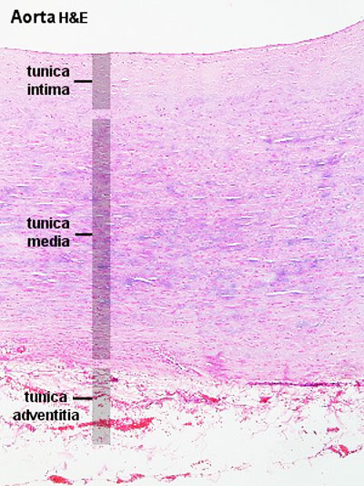
HM Practical - Blood Vessel Histology - Embryology
A Labelled Diagram Of Neuron with Detailed Explanations Diagram Of Neuron. A neuron is a specialized cell, primarily involved in transmitting information through electrical and chemical signals. They are found in the brain, spinal cord and the peripheral nerves. A neuron is also known as the nerve cell. The structure of a neuron varies with their shape and size and it mainly depends upon their ...

Sperm Histology
Two unlabeled slides are viewed under a microscope. Slide A has cells ... Two unlabeled slides are viewed under a microscope. Slide A has cells with a visible nucleus, mitochondria, a cell membrane, a cell wall, and chloroplasts.SlideB has cells with visible nucleus, mitochondria and cell membrane how should the slides be labeled?
_100_04.jpg)
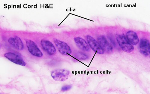
Chapter 7, Page 9 - HistologyOLM
Synapse Diagram Unlabeled - schematron.org Read the definitions, then label the neuron diagram below. axon - the long extension of a neuron that carries nerve impulses away from the body of the cell. axon terminals - the hair-like ends of the axon cell body - the cell body of the neuron; it contains the nucleus (also called the soma. Unlabeled Diagram Of Nervous System.

Plant Cells VS. Animal Cells
Ultrastructural identification of somata and neural processes ... Unlabeled cells were of large, medium and small size. GAD-positive terminals were identified as F1 and F2 types (Guillery's nomenclature) on the basis of their synaptic relations and ultrastructure. Labeled F2 terminals were postsynaptic to retinal (RLP) boutons and presynaptic to unlabeled dendrites in synaptic glomeruli.
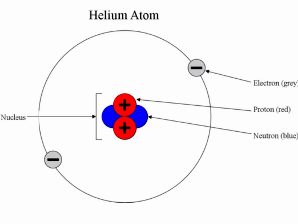
Subatomic Particles by Andrea Cleofas
Packing of DNA into a nucleosome core particle in the nucleus Packing of DNA into a nucleosome core particle in the nucleus Variant Image ID: 74945 Add to Lightbox. Save to Lightbox. Email this page; Link this page ; Print; Please describe! how you will use this image and then you will be able to add this image to your shopping basket. Pricing. Price for. Add To Cart ...
Post a Comment for "42 nucleus unlabeled"