38 ear diagram
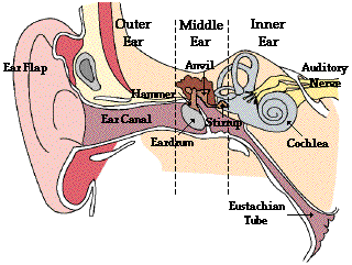
Label Parts of the Human Ear - University of Dayton Label Parts of the Human Ear. Select One Auditory Canal Cochlea Cochlear Nerve Eustachian Tube Incus Malleus Oval Window Pinna Round Window Semicircular Canals Stapes Tympanic Membrane Vestibular Nerve. Select One Auditory Canal Cochlea Cochlear Nerve Eustachian Tube Incus Malleus Oval Window Pinna Round Window Semicircular Canals Stapes ... Well-Labelled Diagram Of Ear With Explanation - BYJUS Diagram of Ear Human ear is a sense organ responsible for hearing and body balance. The outer ear receives the sound waves and transmits them down the ear canal to the eardrum. This causes the eardrum to vibrate and sound is produced. The diagram of ear is important from Class 10 and 12 perspective and is usually asked in the examinations.
Ear Diagram | EdrawMax Editable Templates The ear diagram shows that the ear has external, middle, and inner portions. The outer ear is called the pinna and is made of ridged cartilage covered by skin. Sound funnels through the pinna, the external auditory canal, a short tube that ends at the eardrum. The eardrum is a transparent gray membrane. Attached to the center part of the drum is the middle ear bone.

Ear diagram
Ear Anatomy, Diagram & Pictures | Body Maps - Healthline There are two main sections within the inner ear: the bony labyrinth and the membranous labyrinth. The cochlea, the hearing organ, is located inside the inner ear. The snail-like cochlea is made up... Picture of the Ear: Ear Conditions and Treatments - WebMD The ear has external, middle, and inner portions. The outer ear is called the pinna and is made of ridged cartilage covered by skin. Sound funnels through the pinna into the external auditory... Free Ear Diagram Templates - Edrawsoft Ear Diagram Template Download Template: Get EdrawMax Now! Free Download Popular Latest Flowchart Process Flowchart Workflow BPMN Cross-Functional Flowchart Data Flow Diagram EPC Fault Tree IDEF Diagram Org Chart Basic Org Chart Photo Org Chart Creative Org Chart Family Tree Genogram Network Rack Diagram Network Topology CCTV Network LDAP
Ear diagram. Bones of the Ear - Anatomy Pictures and Information The bones of the ear, also known as the auditory ossicles, are the three smallest bones in the human body. These bones play an important role in the sense of hearing by transmitting sounds to the inner ear. Anatomy. The three auditory ossicles — the malleus, incus, and stapes — are tiny bones found in the middle ear. Ear Anatomy - Outer Ear | McGovern Medical School The outer ear is made up of cartilage and skin. There are three different parts to the outer ear; the tragus, helix and the lobule. EAR CANAL The ear canal starts at the outer ear and ends at the ear drum. The canal is approximately an inch in length. The skin of the ear canal is very sensitive to pain and pressure. Ear - Wikipedia In mammals, the ear is usually described as having three parts—the outer ear, the middle ear and the inner ear. The outer ear consists of the pinna and the ear canal. Since the outer ear is the only visible portion of the ear in most animals, the word "ear" often refers to the external part alone. How to Draw Human Ear Diagram With Labelling #HumanEar Thanks for watching our Channel. how to draw internal structure of human ear,diagram of human ear for class 8,diagram of human ear with labelling,structure o...
PDF the diagram - Central Institute for the Deaf the diagram EAR HOW WE HEAR 1. Sound enters the ear. 2. The ear drum vibrates. 3. The bones in the middle ear move. 4. The fluid inside the cochlea moves. 5. The hair cells inside the cochlea vibrate. 6. The auditory nerve is activated. 7. The message is sent to the brain. Outer Ear Middle Ear Inner Ear Ear Diagram (English} | CID Free Download CONTACT US. 825 S. Taylor Avenue Saint Louis, MO 63110. Toll free: 877.444.4574 Tel: 314.977.0132 Fax: 314.977.0023 Ear Anatomy, Diagram & Structure | What are the Parts of the Ear ... The following ear diagram depicts the inner ear, which contains sensory organs for hearing and balance, and the outer ear, which includes superficial structures. The middle ear is sandwiched... The Ear: Anatomy, Function, and Treatment - Verywell Health Essential organs of human hearing and balance, the ears are located on either side of the head, at the level of the nose. Separated into an inner, middle, and outer ear, each ear is an intricate and complicated mixture of bones, nerves, and muscles. Naturally, these structures are at the heart of hearing loss problems as well as those affecting ...
Blank ear diagrams and quizzes: The fastest way to learn - Kenhub Ear diagrams (labeled and unlabeled) Accelerate your learning with interactive quizzes Sources + Show all Ear anatomy overview Although it's not obvious to look at, the ear is anatomically divided into three portions: External (outer) ear Middle ear Inner ear As you can imagine, there's a lot of associated anatomy to learn for each portion! Ear Diagram - Concha Audiology The cochlea is a fluid-filled organ essential for the transduction of mechanical (vibration) energy to electrical (nerve impulse) energy. Vibrations from the stapes on the oval window cause waves within the fluid, which causes the basilar membrane to move. The movement of the basilar membrane causes a shearing action of hair cells (outer and ... Ear (Anatomy): Overview, Parts and Functions - Biology Dictionary The human ear picks up and interprets high-frequency vibrations of air, while the sound-sensing organs of aquatic animals are designed to pick up high-frequency vibrations in water. Most vertebrates have two ears: one on either side of the head. In some animals, including most mammals, the ear is also used for balance. Human Ear: Structure and Functions (With Diagram) ADVERTISEMENTS: In this article we will discuss about the structure and functions of human ear. Structure of Ear: Each ear consists of three portions: (i) External ear, ADVERTISEMENTS: (ii) Middle ear and (iii) Internal ear. 1. External Ear: It comprises a pinna, external auditory meatus (canal) & tympanic membrane. (i) Pinna: ADVERTISEMENTS: The pinna is […]
Lyric hearing aid: a rare cause of benign necrotising otitis externa ...
Anatomy of the Ear | Inner Ear | Middle Ear | Outer Ear Anatomy of the Ear Anatomy Chart How We Hear Diseases / Abnormalities The ear is made up of three parts: the outer, middle, and inner ear. All three parts of the ear are important for detecting sound by working together to move sound from the outer part through the middle and into the inner part of the ear. Ears also help to maintain balance.

CGArena : Modeling Ears in 3D
Inner Ear Diagram Photos and Premium High Res Pictures - Getty Images human ear 108 Inner Ear Diagram Premium High Res Photos Browse 108 inner ear diagram stock photos and images available, or search for human ear to find more great stock photos and pictures. of 2 NEXT

Face lift tutorial, Scar pitfalls: page 8 - FacialSurgery.com
Ear anatomy: Parts and functions - Kenhub The ear is a complex part of an even more complex sensory system. It is situated bilaterally on the human skull, at the same level as the nose. The main functions of the ear are, of course, hearing, as well as constantly maintaining balance. The ear is anatomically divided into three portions: External ear Middle ear Internal ear

Ear Labeling
The ear canal: Anatomy, diagram, and common conditions The ear has outer, middle, and inner portions. The ear canal and outer cartilage of the ear make up the outer ear. The ear canal transports sound from the outer ear to the eardrum, which is in the...
Openphysics/Sound - WikiEducator
Human Ear Diagram - Bodytomy The Structure of Human Ear. Helix: It is the prominent outer rim of the external ear. Antihelix: It is the cartilage curve that is situated parallel to the helix. Crus of the Helix: It is the landmark of the outer ear, situated right above the pointy protrusion known as the tragus. Auditory Ossicles: The three small bones in the middle ear ...

Pin by Logan on fashion | Cool ear piercings, Ear piercing diagram, Ear ...
Ear Anatomy: Understanding the Outer, Middle, and Inner Parts of the Ear The external auditory meatus, or ear canal, is a narrow canal that leads from the concha to the tympanic membrane, or eardrum. Sound waves are delivered through this canal. This canal is prone to ear infections. Tragus This is the small, rigid part of the ears along the front of the ear, adjacent to the face.

Axial skeleton - Isaiah's Anatomy Website
Home - Embibe Exams we bring you the latest exam news and scoring secrets click below to select your exam
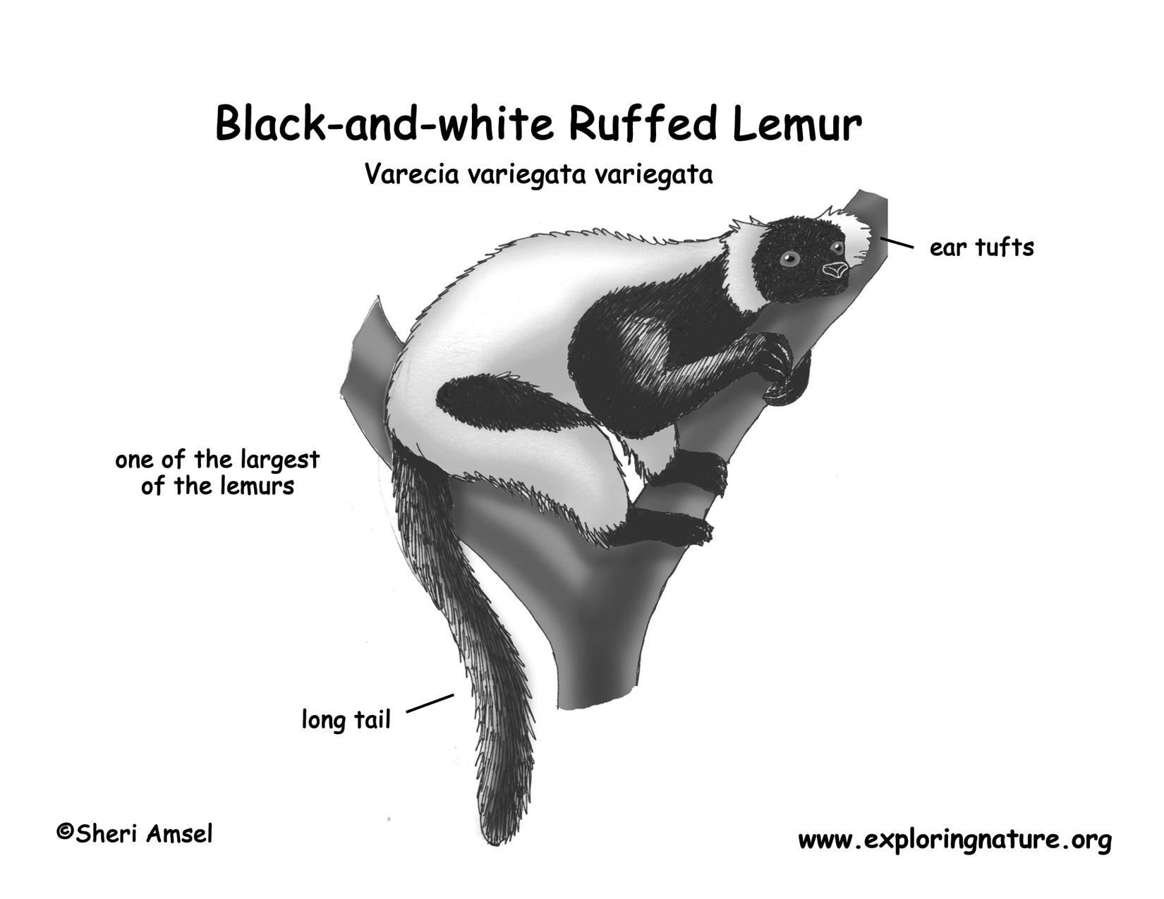
Lemur (Black-and-white Ruffed)
Human Ear Anatomy - Parts of Ear Structure, Diagram and Ear Problems Human ear The ear is divided into three anatomical regions: the external ear, the middle ear, and the internal ear (Figure 2). The external ear is the visible portion of the ear, and it collects and directs sound waves to the eardrum. The middle ear is a chamber located within the petrous portion of the temporal bone.

67 Unique Ear Piercing Ideas That You Never Thought About
Ear Canal Diagram, Pictures & Anatomy | Body Maps Ear Canal Diagram, Pictures & Anatomy | Body Maps Human body Head Ear Ear canal External acoustic meatus The ear canal, also called the external acoustic meatus, is a passage comprised of bone and...
Post a Comment for "38 ear diagram"